ENDOSCOPIC DECOMPRESSION IN LUMBAR
DEGENERATIVE FORAMINAL STENOSIS
(FORAMINOPLASTY)
Eric GOZLAN, Paris; Benoit LAVIGNOLLE, Bordeaux (France)
ISMISS, Zurich- Jan. 2009
PURPOSE:
To evaluate the efficacy and safety of transforaminal endoscopic decompression in cases of lumbar degenerative foraminal stenosis.
METHOD:
Inclusion criteria
Symptomatology of monoradicular and unilateral claudication resisting from over 6 months of conservative treatment and particularly foraminal injections of positive, but no lasting effect. Their aim is to select the right symptomatic nerve root and the pathologic foramen.
Necessity of imagery being concordant with symptomatic nerve root with a clear unilateral foraminal stenosis. Presence, eventually of a Grade I spondylolisthesis
Initial Evaluation
From June 2005 to March 2007, 201 patients were admitted in this study and evaluated at 0.1 and 3 months (201 cases) and 12 months (117 cases)
Sex ratio was M/F = 85/116, mean age 53.3 years (min=26, max=82).
Average duration of symptoms was 4.32 years for recurrent cases and 9.5 months for consecutive evolutionary cases.
67 out of 201 presented neurological signs.
52 patients had already had primary surgery and 8 chemonucleolysis.
Also, Foraminoplasty has been performed at 2 levels in 18 cases.
RESULTS:
- At 3 months post operatively we noted that:
Neurological signs had disappeared in 67% of the cases.
Radicular pain was reduced on average by 72% on the AVS and lumbar pain by 57%
The DALLAS score was reduced by 46,5%
Also, 81% of the patients considered the result as a success:
- At 12 months post operatively (117 patients) the results were:
Radicular pain decreased on average by 75% and 64% for lumbar pain.
The DALLAS score reduced on average by 54%.
We noted that walking distance had significantly improved at least over 1 km, in the successes.
The success rate increased to 89%.
- No major complications appeared in this study.
DISCUSSION:
Transforaminal Endoscopic Discectomy treating discal herniations is a technique fully validated.
It can be extended to degenerative foraminal stenosis associated or not to a herniation with minimally invasive conditions.
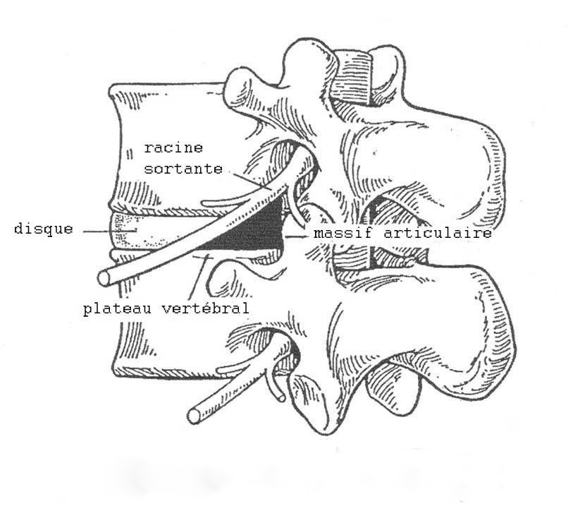
This technique allows us to visualize easily the exiting nerve root in the foramen as well as the articular process.
It is then possible to enlarge the foramen with trephines and laser and also decompress the disk.
CONCLUSION:
This study has proved statistically significant good results at 3 months and even better ones at 12 months.
This technique allows a minimal transforaminal access without risk of fibrosis and destabilization